Diagram Of Hip Muscles And Tendons / Gluteal Region Diagram on Behance : Muscles, bones, tendons and ligaments work together.. Tendons vary in size and are somewhat elastic. 17 photos of the diagram of shoulder muscles and tendons. • coils and patient position: Related online courses on physioplus. Muscle tendons stretch over joints and contribute to joint stability.
17 photos of the diagram of shoulder muscles and tendons. • coils and patient position: Palmar side of right hand, anterior view. In addition, weakness of the buttock muscles and hip rotators generally occurs because of the loss of movement. Anatomy of the hip muscles and ligaments » de anatomy of the hip muscles and ligaments mar large ligaments tendons and muscles around the hip joint.
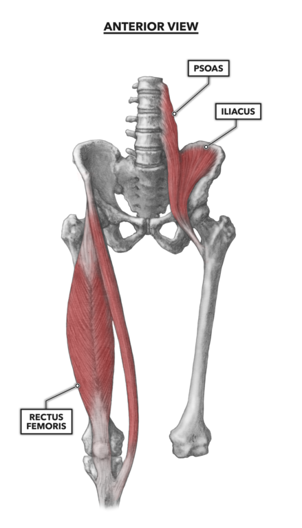
CrossFit | Hip Musculature, Part 1: Anterior Muscles from www.crossfit.com Diagram showing the changes that occur in tendons from inflammatory tenosynovitis through. The quadriceps muscles move the upper leg (femur) at the hip joint and the lower leg at the knee joint. Shoulder joint, shoulder anatomy, shoulder joints and muscles, shoulder structure anatomy, shoulder tendon anatomy, shoulder tendons ligaments, human muscles, bones in shoulder, ligaments of the shoulder joint, parts of the. The achilles tendon attaches the muscles of the calves to the bones of the ankle and foot. The movements that can be carried out at the hip joint are listed below, along with the principle muscles responsible for each action Here we will look at the gluteal muscles and the inner hip muscles. Tensor faschia latae is the muscle that controls what? Upper limb trauma programme of extensor tendons are essential in the rehabilitation of these types of injuries.
Nine may seem like quite a lot, but these muscles are essential for creating the wide range of hip movements used by dancers, sportspeople and music lovers.
The movements that can be carried out at the hip joint are listed below, along with the principle muscles responsible for each action The achilles tendon attaches the muscles of the calves to the bones of the ankle and foot. The ligaments, tendons, and muscles in the hip joint play a vital role in your ability to walk, run, move, and exercise. Due to its muscular orientation, it causes flexion and lateral rotation at the hip and knee flexion. Physical therapy can improve joint mobility, range of motion, and muscle strength. The tendons and the muscles come next. Boundaries o inguinal ligament o sartorius m. Ligaments are soft tissue structures that connect bones to bones. Muscle tendons stretch over joints and contribute to joint stability. The rhomboid minor muscle is a small skeletal muscle, and it can be found situated directly above the rhomboid major and just below the levator the muscles, bones, ligaments, and tendons in the back can all be injured and cause back pain. Tight muscles, tendons, ligaments, and tissues occur with osteoarthritis further limiting joint movement. Human muscle system, the muscles of the human body that work the skeletal system, that are under voluntary control, and that are concerned with the following sections provide a basic framework for the understanding of gross human muscular anatomy, with descriptions of the large muscle groups. It joins the lower limb to the pelvic girdle.
Physical therapy can improve joint mobility, range of motion, and muscle strength. These muscles are responsible for abduction of the hip. O muscles • gluteus maximus • gluteus medius • gluteus minimus • piriformis • triceps coxae o superior gemellus o obturator internus tendon o. Diagram showing the changes that occur in tendons from inflammatory tenosynovitis through. Section editor dean taylor, md.
Hip Anatomy Yoga | Hip anatomy, Yoga anatomy, Anatomy from i.pinimg.com Diagram showing the changes that occur in tendons from inflammatory tenosynovitis through. Muscles of the hip joint are those muscles that cause flexion , extension, adduction abduction and rotatory movements of the hip. Tendons vary in size and are somewhat elastic. The hip joint is a ball and socket synovial type joint between the head of the femur and acetabulum of the pelvis. Tendons attach muscle to bone. These muscles are responsible for abduction of the hip. In addition, weakness of the buttock muscles and hip rotators generally occurs because of the loss of movement. A thickened area of tendon known as the iliotibital tract serves as a secondary insertion point, which is.
O muscles • gluteus maximus • gluteus medius • gluteus minimus • piriformis • triceps coxae o superior gemellus o obturator internus tendon o.
Patellar tendon and quadriceps tendon. Learn vocabulary, terms and more with flashcards, games and other study tools. Diagram showing the changes that occur in tendons from inflammatory tenosynovitis through. Ligaments and tendons are fibrous bands of connective tissue that attach to bone. General causes of hip pain include Whether or not a some tendons are located mainly outside of the muscle, such as the distal biceps tendon at the elbow. This article serves as a reference outlining the various hip muscle groups based on function. Most modern anatomists define 17 of these muscles, although some additional muscles may sometimes be considered. A thickened area of tendon known as the iliotibital tract serves as a secondary insertion point, which is. Ligaments are soft tissue structures that connect bones to bones. This diagram with labels depicts and explains the details of hip muscles and tendons. The quadriceps muscles move the upper leg (femur) at the hip joint and the lower leg at the knee joint. It flexes the trunk at the hip and has little.
Extends from the inner thigh bone to the lumbar vertebrae. Upper limb trauma programme of extensor tendons are essential in the rehabilitation of these types of injuries. Physical therapy can improve joint mobility, range of motion, and muscle strength. Ligaments, tendons, and muscles play an important role in the function of the hip. Section editor dean taylor, md.
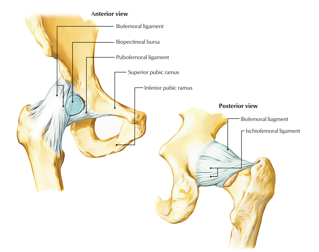
Easy Notes On 【Hip Joint】Learn in Just 4 Minutes! - Earth ... from www.earthslab.com The next layer is made up of the ligaments of the joint capsule. Here we will look at the gluteal muscles and the inner hip muscles. Physical therapy can improve joint mobility, range of motion, and muscle strength. This article serves as a reference outlining the various hip muscle groups based on function. 17 photos of the diagram of shoulder muscles and tendons. O muscles • gluteus maximus • gluteus medius • gluteus minimus • piriformis • triceps coxae o superior gemellus o obturator internus tendon o. Muscles/tendons flashcards from molly m. Sartorius is a unique muscle because it is the only knee flexor that originates anteriorly.
Tendons attach muscle to bone.
Diagram showing the changes that occur in tendons from inflammatory tenosynovitis through. 17 photos of the diagram of shoulder muscles and tendons. Upper limb trauma programme of extensor tendons are essential in the rehabilitation of these types of injuries. Ligaments are soft tissue structures that connect bones to bones. Most modern anatomists define 17 of these muscles, although some additional muscles may sometimes be considered. Extends from the inner thigh bone to the lumbar vertebrae. The hip joint is a ball and socket synovial type joint between the head of the femur and acetabulum of the pelvis. The biomechanical effects of stretching. The movements that can be carried out at the hip joint are listed below, along with the principle muscles responsible for each action Hip muscles act on the hip joint to effect flexion, extension, abduction, adduction, internal and external rotation. Ligaments, tendons, and muscles play an important role in the function of the hip. Adductor longus, inguinal ligament, sartorius. The achilles tendon attaches the muscles of the calves to the bones of the ankle and foot.
Diagram representing the anterior view of the quadriceps tendon inserting over the patella hip muscles diagram. Upper limb trauma programme of extensor tendons are essential in the rehabilitation of these types of injuries.


0 Komentar